
✅ Medically reviewed
A knee MRI scan uses a strong magnet and radio waves. It creates detailed images of cartilage, ligaments, tendons and bone. Knee problems are widespread in the UK. Arthritis UK, formerly Versus Arthritis, reports that over 10 million people in the UK have arthritis. An estimated 5.4 million have knee osteoarthritis. NHS England also recorded around 4.9 million MRI tests in the year to March 2025.
Doctors request a knee scan when an X-ray cannot explain swelling, locking or lasting pain. The machine knocks like an enthusiastic woodpecker, yet the experience is calmer than its reputation suggests. The test uses no radiation and is painless.
What Does a Knee MRI Show?
A knee MRI builds slice-by-slice pictures of soft tissue and bone. The magnet lines up water molecules inside the body. Radio pulses then knock them out of line. A computer converts the returning signals into images from three angles.
The images show structures that plain X-rays cannot display.
Structures the Images Reveal
Radiologists examine the menisci, the cruciate and collateral ligaments, tendons, cartilage and bone marrow. Fluid inside the joint appears clearly. So does swelling within the bone, which often follows a bruise or stress injury. Fluid-filled swellings, including a cyst behind the knee, also stand out.
What Radiologists Look For
Radiologists check cartilage thickness, tear patterns and bone swelling. They also look for loose fragments, excess fluid and signs of infection. Every finding goes into a written report for the referring doctor. A knee MRI scan therefore answers several questions in a single appointment.
Why Scanner Strength Matters
Most hospital scanners run at 1.5 or 3 tesla. The higher strength usually gives sharper images in less time. Both types diagnose common knee injuries reliably. Open scanners suit patients with severe anxiety or a larger frame, though image detail may be lower.
Knee MRI vs X-Ray and Which Test Suits Which Problem
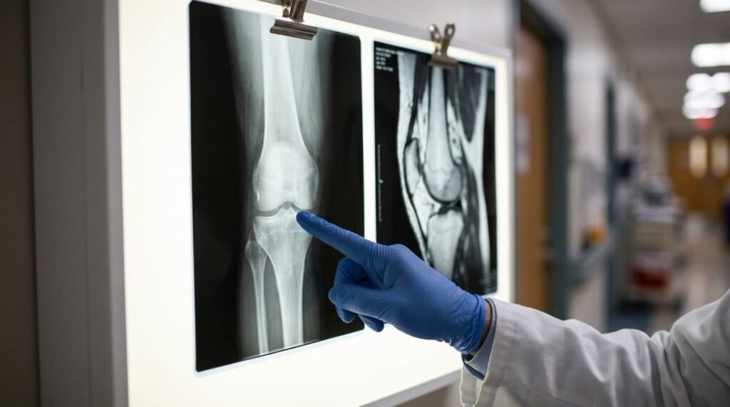
X-rays remain the first test for most knee complaints. They show bone shape, fractures and the gap between joint surfaces. That gap narrows as cartilage wears away. Doctors therefore use X-rays to assess knee osteoarthritis quickly.
MRI adds the soft tissue detail that X-rays lack. It shows a torn ligament, a damaged meniscus or early cartilage loss. It also reveals a bone bruise that hides on plain films.
How Ultrasound and CT Compare
Ultrasound examines tendons and fluid in real time, but sound waves cannot see deep inside the joint. CT shows fine bone detail and suits complex fractures. Neither matches MRI for ligament and cartilage assessment. Ultrasound also guides many joint injections.
Each test answers a different question, so doctors choose by symptoms rather than habit.
When Doctors Order a Knee MRI Scan
A doctor requests the test when symptoms point to a soft tissue problem. The X-ray has usually come back normal or unclear by then.
Common Knee MRI Uses
Typical reasons include persistent swelling, a knee that locks or gives way, and pain after a twisting injury. Surgeons also use MRI before arthroscopy and after surgery to check healing. Doctors occasionally order it to investigate infection or a suspected bone tumour. Athletes and manual workers often need clear answers quickly, since a tear can limit work and sport.
MRI Knee Meniscus Tear Detection
The menisci are two C-shaped cartilage pads that cushion the joint. A twist while the foot stays planted often tears one. The images show the location and pattern of the tear. That detail helps surgeons choose between rehabilitation and an operation.
MRI Knee Ligament Tear Assessment
The anterior cruciate ligament, posterior cruciate ligament and collateral ligaments keep the joint stable. Sports injuries and road accidents commonly damage them. MRI separates a partial tear from a complete rupture. That distinction changes treatment decisions.
After Injury or Surgery
A knee MRI scan after surgery checks that a repaired ligament or meniscus is healing. It also looks for scar tissue, fluid and infection. Metal implants distort the picture, so pain after a knee replacement often starts with X-rays instead.
When a Scan Adds Little
Advanced arthritis usually needs no MRI. NICE guidance supports diagnosing osteoarthritis from symptoms and examination in most adults aged 45 or over. A standing X-ray confirms the picture when needed. Patients with mild to moderate osteoarthritis sometimes consider an Arthrosamid injection. Clinicians judge suitability through history, examination and imaging where relevant.
How to Prepare for a Knee MRI
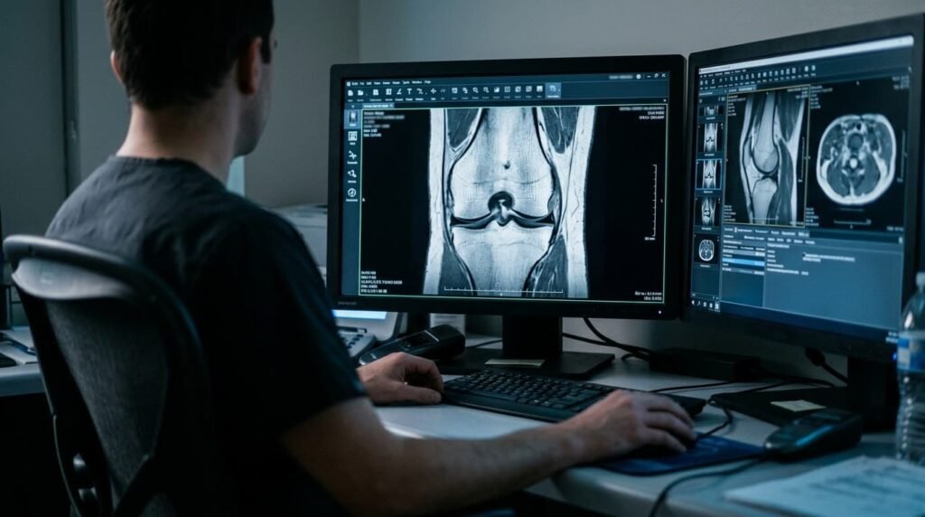
Preparation is short and simple. Most centres allow normal eating, drinking and medication unless the appointment letter says otherwise. Patients complete a safety questionnaire on arrival. The form asks about previous operations, earlier scans and any metal injuries, such as grinding fragments from workshop accidents. Accurate answers protect safety. Arriving early avoids any rush.
What to Wear to a Knee MRI
Loose, comfortable clothing without metal works best. Zips, buttons, underwired items and sportswear with metallic thread can interfere with the magnet. Many departments supply a gown. Shorts or loose trousers that roll above the knee also work well. Jewellery, watches and hairpins stay at home. The magnet will not be impressed by a belt buckle.
Metal, Devices and Knee MRI Implants
Metal is the main safety issue because the magnet stays switched on at all times. Patients must tell the radiographer about pacemakers, cochlear implants, insulin pumps, aneurysm clips and metal fragments. Pregnancy, kidney disease, and past reactions to contrast dye also need to be mentioned.
Most orthopaedic implants, including knee replacements, are safe to scan. They can blur the picture near the metal. Radiographers sometimes adjust the settings to reduce that distortion.
Video explainers on joint pain and arthritis care appear on the Dr SNA Clinic YouTube channel.
Questions Worth Asking Beforehand
Patients can raise several practical points with the referring team. Useful topics include whether contrast will be used, how long the appointment lasts and whether sedation needs a lift home. A knee MRI scan runs smoothly when staff know every detail in advance.
Knee MRI: What to Expect on the Day
A knee MRI scan feels like lying still on a firm table in a loud room. Nothing touches the knee except a padded frame. The magnet produces no sensation at all.
Step-by-Step Through the Appointment
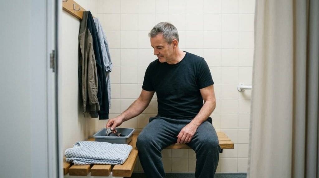
The radiographer checks the safety form and positions the patient on a padded table. A coil, which is a padded frame that detects the signal, wraps around the knee. Straps and cushions hold the leg still. Earplugs or headphones follow, and many centres offer music.
The table then slides into the scanner feet first. Only the lower body enters the tunnel, so the head and chest usually stay outside. The radiographer watches from the next room and can talk to the patient between sequences. A call button stays within reach throughout.
How Long Does a Knee MRI Take?
Most knee scans last 20 to 45 minutes. NHS trusts quote 15 to 90 minutes for MRI in general, depending on the body area. Contrast dye adds time. Movement also adds time, because the radiographer repeats any blurred sequence. Patients should plan for about an hour in total, including check-in and screening.
Is a Knee MRI Painful?
No. The magnet causes no sensation, and the test involves no radiation. Any discomfort comes from lying still rather than from the scan itself. A pillow under the knees or a change of position often helps. Patients who receive contrast feel only the pinch of the needle.
Knee MRI Claustrophobia and How to Manage It
NHS trust leaflets suggest that about 1 per cent of patients feel claustrophobic during a scan. A knee study is usually easier, since the head stays outside the tunnel. Patients with anxiety should tell the doctor before the appointment. Options include a mild prescribed sedative, a wide-bore scanner, calming music and a chat with the radiographer. Some departments allow a visit to the scanner room before the appointment. Closing the eyes before entering and keeping them closed also helps many patients.
Patients who take a sedative must arrange a lift home.
Tips for Staying Still
Stillness matters more than anything else during the appointment. Breathing normally is fine, and the muscles can relax. Counting breaths or planning the weekend helps pass the time. A knee MRI scan rewards the calm patient with sharper images and fewer repeats. Coughing or fidgeting blurs the pictures, so a tickle in the throat deserves a mention beforehand.
Knee MRI With or Without Contrast
Most knee scans need no contrast. Standard sequences show meniscus, ligament and cartilage injuries clearly. Doctors add contrast when inflammation, a tumour or a joint infection is suspected.
The dye is a gadolinium-based agent given through a vein in the arm. It can cause a brief metallic taste or a wave of warmth. Radiographers check kidney function first when needed. Allergic reactions are rare and usually mild.
What Is an MR Arthrogram Knee Study?
An MR arthrogram injects dilute contrast directly into the joint before scanning. The fluid outlines cartilage and meniscus edges more sharply. Surgeons often request it after a previous meniscus repair. The procedure takes longer than a standard scan.
Knee MRI Accuracy, Risks and Safety
Knee MRI Accuracy for Meniscus and Ligament Injuries
Research supports the reliability of the test. A 2026 pooled analysis of 75 studies reported 91 per cent sensitivity for medial meniscus tears. Sensitivity measures how often the test finds a real tear. The same analysis reported 94 per cent specificity for lateral meniscus tears. Image quality, scanner strength and radiologist experience all influence the final result.
Knee MRI Risks and Safety Checks
The magnetic field itself is harmless. Risks arise mainly from metal devices and contrast dye. Allergic reactions are uncommon. Nephrogenic systemic fibrosis, a kidney-related complication, is exceptionally rare with modern agents. Staff monitor vital signs when sedation is used. Very small amounts of gadolinium can remain in the body after repeated scans, though no health effects are known.
Who Should Delay the Test
Some pacemakers and implanted devices are unsafe in the magnet, so staff screen every patient. Doctors may postpone non-urgent scans during pregnancy. Gadolinium dye is generally avoided in pregnancy except in specific cases. Patients with kidney disease need blood tests before contrast.
Why Abnormal Findings Do Not Always Explain Pain
One study of 230 pain-free adults found abnormalities in 97 per cent of knees. Meniscal tears appeared in around 30 per cent of them. A report therefore needs clinical context before anyone acts on it. Doctors treat the patient, not the picture.
Pain may also start outside the knee. Knee pain from hip problems is a common example, and a hip examination often settles the question.
Knee MRI Results: How Long Reports Usually Take

A radiologist studies the images and writes a report. The report goes to the doctor who requested the test. Reports often arrive within a few days, though some NHS leaflets quote up to two weeks. Patients who have heard nothing after two weeks should contact the referring team.
Patient portals sometimes release results before a clinician explains them. Terms such as tear, degeneration and effusion can sound alarming in isolation. A report is one piece of evidence, not a verdict.
Clinicians combine the findings with symptoms, examination and X-rays. The result then guides the next step, whether rehabilitation, an injection or surgery. Non-surgical orthopaedic treatment often forms the first stage, and the plan changes as joint damage advances.
Aftercare and Next Steps
Patients dress and leave straight after the scan. No recovery period applies unless sedation was given. The kidneys clear gadolinium dye, so most patients resume normal activity immediately. A follow-up appointment then explains the findings and the options. A knee MRI scan result guides the plan but never replaces a conversation with the treating clinician.
Frequently Asked Questions
How long does a knee MRI take when contrast is added?
Contrast usually extends the appointment by several minutes. The radiographer must place a needle in a vein and run extra sequences. Patients should still plan for roughly an hour in the department. The radiographer confirms the timing before the scan starts.
Can patients with metal implants have a knee MRI?
Most orthopaedic implants, including knee replacements, are safe in the scanner. The radiographer needs to know about every device and previous operation. Metal can blur the image near the implant. Centres often adjust the settings to limit that effect.
Does an MRI knee meniscus tear always need surgery?
No. Many tears settle with physiotherapy, activity changes and pain relief. Surgeons consider the tear pattern, the symptoms and the patient’s age. Locking or a stuck joint raises the case for an operation.
What does a knee MRI show that ultrasound misses?
Ultrasound cannot see through bone, so deep joint structures stay hidden. MRI shows the cruciate ligaments, the menisci and the cartilage surface in detail. It also reveals bone marrow swelling. Ultrasound still suits tendons and superficial fluid.
How soon do patients get the results?
Reports often reach the referring doctor within a few days. Some departments take up to two weeks. The referring doctor then explains the findings. Patients should ask about the expected timescale at the appointment.
A knee MRI scan gives clear, radiation-free images of the soft tissues that X-rays miss. Preparation is simple, the appointment is painless, and reports usually follow within days. Findings carry the most value when clinicians read them alongside symptoms and examination.
Mr Syed Nadeem Abbas is a non-surgical knee and joint pain specialist. Patients with ongoing knee pain can book a consultation to discuss the right diagnostic steps.
Read more: A Knee Pain Doctor Helps Identify The Right Treatment Before Symptoms Worsen
Read more: Knee Pain Treatment Options That Actually Relieve Discomfort


