Cysts Behind the Knee Can Signal Several Different Joint Conditions

✅ Medically reviewed | Updated september 2026
Cysts behind the knee are a common but often misunderstood finding in orthopaedic practice. Many patients assume any swelling in this area is a Baker’s cyst. In reality, several distinct conditions can produce a similar lump. Some originate from the joint capsule itself, while others develop within nearby tendons or a torn meniscus. Distinguishing between these types matters, because treatment differs significantly from one cause to another. Age, activity level, and existing joint health all influence which type is most likely. The following sections outline the main types found behind the knee, their causes, and when medical review becomes necessary.
What Counts as a Cyst Behind the Knee
A cyst is a closed sac containing fluid, semi-fluid, or gel-like material. Behind the knee, this sac usually forms from excess synovial fluid, the natural lubricant inside the joint capsule. Swelling can range from barely noticeable to a firm, tender mass that limits movement. Location often provides the first clue to type. A lump sitting directly behind the joint line frequently traces back to the joint capsule itself. One appearing slightly to the side may originate from a meniscal tear or a tendon sheath.
Pain levels vary widely between patients. Some notice only mild tightness after long walks. Others experience persistent discomfort that worsens with bending. Reduced knee movement and a visible lump remain the most reported signs across cyst types. Skin colour and temperature over the swelling usually stay normal, which helps rule out infection at first glance. A firm, well-defined edge is typical, whereas a spreading or poorly defined margin deserves closer attention. A fluid-filled lump behind the knee should always prompt clinical assessment rather than self-diagnosis. Appearance alone rarely confirms the exact cause, and two very different conditions can look almost identical to the naked eye. A short delay in seeking review rarely causes harm. An extended delay, however, can allow an underlying joint problem to worsen unnoticed over time.
Types of Cysts Behind the Knee
Several distinct conditions can present as a lump behind the knee. Each has its own cause, typical location, and management pathway. Recognising the differences helps set realistic expectations for treatment.
Popliteal (Baker’s) Cyst
The most recognised type is the popliteal cyst, commonly known as a Baker’s cyst. It develops when synovial fluid herniates into the bursa behind the knee. Osteoarthritis and meniscal tears are frequent underlying triggers, since both increase joint fluid production over time. Detailed causes, symptoms, and treatment routes for this specific condition are covered separately in dedicated clinical guidance. Within this broader discussion, a Baker’s cyst represents just one possible explanation for posterior knee swelling. It is not the automatic default diagnosis simply because it is the best-known one.
Meniscal Cyst
A meniscal cyst forms when synovial fluid collects around a torn meniscus. Unlike a Baker’s cyst, this type tends to sit closer to the joint line rather than directly behind it. Pain frequently worsens during twisting movements or deep knee bending. Mechanical symptoms, such as catching or locking, often accompany a meniscal cyst and rarely appear with a straightforward Baker’s cyst. These features help distinguish it from cysts unrelated to meniscal damage.
Ganglion Cyst Knee
A ganglion cyst knee presentation typically arises from a tendon sheath, ligament, or joint capsule. These cysts contain thick, gel-like fluid rather than the thinner synovial fluid seen in a Baker’s cyst. Some remain small and symptom-free for years. Others enlarge gradually and press on nearby structures, causing localised discomfort or mild nerve irritation. This type can appear at the front, side, or back of the knee joint, unlike posterior-only presentations.
Semimembranosus Bursa Cyst
The semimembranosus bursa cyst develops in a small fluid sac near the inner side of the knee. This location sits close to the hamstring tendon. It is sometimes confused with a Baker’s cyst because of its similar posterior position. However, its origin lies in a natural bursa rather than the joint capsule directly. Overuse and repetitive knee bending are commonly linked to this specific type. Physically active adults who kneel or squat often at work face higher exposure.
Synovial Cyst Knee
A synovial cyst knee presentation shares features with several other types, since it also originates from the joint lining. Chronic inflammation, whether from osteoarthritis or an autoimmune condition, can drive excess fluid production well beyond normal levels. Over time, this fluid may track into surrounding tissue and form a distinct cyst wall. Imaging is often required to confirm this specific diagnosis and separate it from a straightforward Baker’s cyst.
Common Causes of a Cyst Behind the Knee
Multiple underlying joint problems sit behind most cases seen in clinic. Osteoarthritis remains one of the most frequent triggers, since joint wear increases synovial fluid production steadily over months. Meniscal tears, particularly those affecting the posterior horn, are strongly associated with fluid build-up behind the joint. Rheumatoid arthritis and other inflammatory joint conditions can also drive excess synovial fluid production well beyond normal ranges. The mechanism behind rheumatoid arthritis affecting the knee joint over time explains this inflammatory process in more depth.
Repetitive strain from certain sports or occupations may irritate the joint capsule over months or years. Kneeling occupations and repetitive squatting both increase bursa irritation near the back of the joint. Previous knee injuries, even those treated years earlier, sometimes resurface as delayed cyst formation. Gout and other crystal-related joint conditions occasionally contribute to excess fluid production as well. Age plays a role too, since joint wear and synovial fluid changes both become more common after forty. Identifying the root cause matters more than treating the visible lump alone. Without addressing the underlying joint problem, a drained or removed cyst frequently returns within months, sometimes larger than before.
Is It a Baker’s Cyst or Something Else
Patients often ask a simple but important question: is it a Baker’s cyst or something else entirely? Several clues help separate the two. A Baker’s cyst sits centrally behind the knee and often becomes more visible when the leg is fully straightened. Other cyst types may sit slightly off-centre or closer to a specific tendon or ligament. The onset pattern also differs between conditions. A Baker’s cyst usually develops gradually alongside existing joint problems such as arthritis. Meniscal or ganglion cysts sometimes appear more suddenly, particularly following a twisting injury.
Lump Behind Knee Not Baker’s Cyst: Key Differences
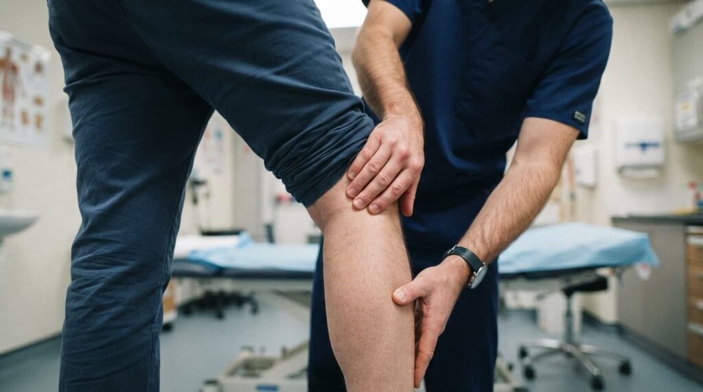
A lump behind the knee not of Baker’s cyst in origin often behaves differently under movement. Meniscal cysts tend to firm up during knee extension and soften slightly during flexion. Ganglion cysts frequently feel more rubbery and mobile under the skin than a typical fluid-filled sac. Semimembranosus cysts sit slightly more medial than a classic Baker’s cyst location. None of these distinctions replaces a proper clinical examination. However, they help explain why self-diagnosis carries a genuine risk of delayed treatment. This risk grows when a lump changes shape or size over a short period.
When to Worry About a Lump Behind the Knee
Most cysts behind the knee are benign and manageable with straightforward treatment. However, certain signs warrant urgent medical attention rather than routine review.
Popliteal Artery Aneurysm: A Rare but Serious Mimic
A popliteal artery aneurysm can occasionally mimic a simple cyst on first inspection. This condition involves abnormal widening of the main artery running behind the knee. Unlike a fluid-filled cyst, it carries a pulsatile quality that a clinician can detect on careful examination. Risk increases with age, smoking history, and existing vascular disease elsewhere in the body. Left untreated, this rare condition can lead to serious circulation problems affecting the whole leg. Sudden onset of a firm, pulsating lump should always receive same-week medical assessment rather than a wait-and-see approach.
Baker’s Cyst vs Blood Clot
Understanding the difference between a Baker’s cyst and a blood clot can be genuinely lifesaving. A ruptured Baker’s cyst can release fluid into the calf, causing sudden pain and swelling. This presentation closely resembles deep vein thrombosis on first appearance. Redness, warmth, and calf tenderness following a burst cyst should never be dismissed as routine bruising. Emergency assessment rules out clot formation quickly and safely. The two conditions require entirely different management pathways, and neither should be assumed without proper testing.
Knee Cyst Diagnosis
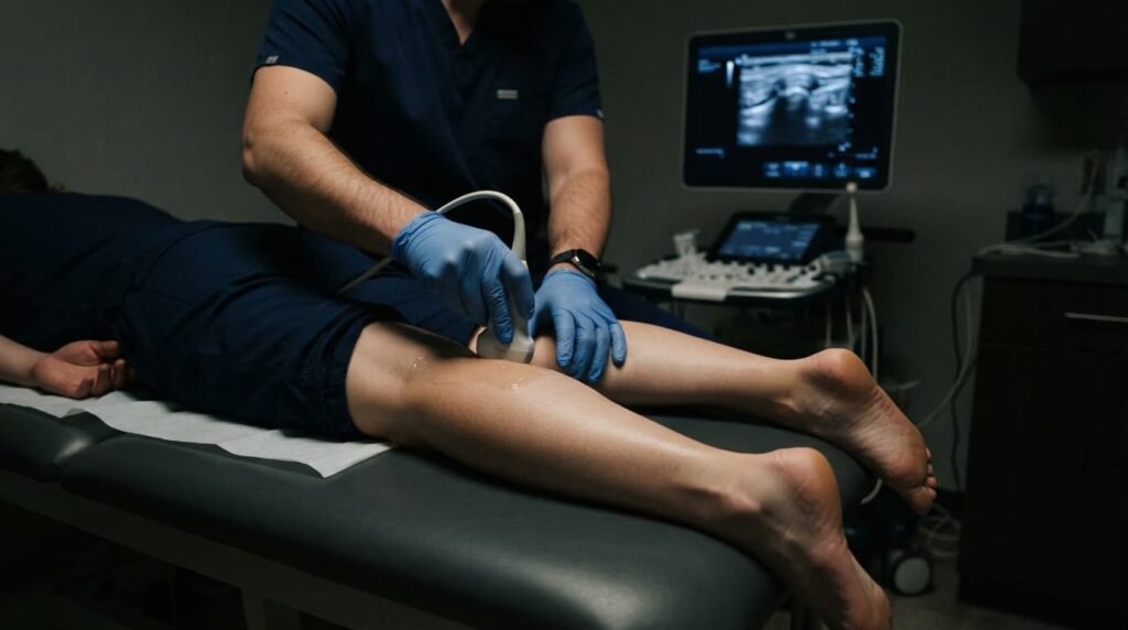
Accurate diagnosis relies on a combination of history-taking, physical examination, and targeted imaging. Ultrasound remains a first-line tool, since it clearly shows fluid-filled structures and their relationship to nearby tissue. It also distinguishes a solid mass from a genuine fluid collection quickly and without radiation. MRI provides more detail when a meniscal tear or ganglion cyst is suspected underneath the visible swelling. It also maps the exact size and depth of the cyst before any procedure. Blood tests may be requested when an inflammatory or autoimmune cause seems likely. Rheumatoid arthritis often prompts this testing, alongside other joint symptoms such as stiffness or morning pain.
Aspiration involves withdrawing a small fluid sample with a fine needle. This step can confirm the diagnosis while relieving pressure at the same time. Fluid analysis occasionally reveals infection or crystal deposits that change the treatment plan entirely. A short period of observation sometimes follows aspiration to confirm the swelling has genuinely settled.
Knee Cyst Treatment Options
Treatment depends entirely on the underlying cause rather than the visible cyst itself. Conservative measures, including rest, compression, and anti-inflammatory medication, often reduce symptoms for smaller presentations. Physiotherapy strengthens surrounding muscles and can ease pressure on an irritated joint capsule over several weeks. A structured exercise programme also improves knee stability, which reduces the chance of recurrence.
Aspiration removes fluid directly but does not address the root cause on its own. Recurrence remains common without further treatment aimed at the underlying joint problem. Steroid injections sometimes accompany aspiration to reduce local inflammation for a longer period. Addressing an underlying meniscal tear or degenerative joint condition offers a genuinely lasting solution rather than temporary relief. Surgical removal is reserved for larger, persistent, or repeatedly recurring presentations that fail conservative care. A cyst treated without fixing its underlying cause tends to return within months. Most patients notice steady improvement within six to twelve weeks once the underlying joint issue receives proper attention.
Living With a Cyst Behind the Knee
Simple self-care measures can ease symptoms while awaiting a full clinical assessment. Gentle rest, avoiding deep squatting, and elevating the leg periodically all help reduce pressure. A supportive bandage may limit swelling without restricting circulation when fitted correctly. Ice application for short periods can calm acute discomfort after activity. Gradual, low-impact movement, rather than complete immobility, generally supports better long-term joint function.
Daily activities rarely need to stop completely, though high-impact exercise is best paused until review. Low-impact options, such as swimming or gentle cycling, often remain comfortable during this period. Any sudden change in size, colour, or pain level should prompt immediate reassessment rather than continued self-management. Keeping a simple note of symptom changes can also help a clinician track progress across appointments. Bringing this record to a review appointment often speeds up decision-making around further imaging or treatment adjustments.
Frequently Asked Questions
What is the most common cause of cysts behind the knee?
Osteoarthritis and meniscal tears account for most cases, since both increase synovial fluid production within the joint.
Can a knee cyst disappear without treatment?
Small cysts sometimes shrink once the underlying joint problem improves, though larger cysts usually require active management.
How is a knee cyst diagnosis usually confirmed?
Ultrasound and MRI remain the standard tools, sometimes supported by blood tests when an inflammatory cause is suspected.
Is a lump behind the knee not Baker’s cyst in origin dangerous?
Not usually, though ganglion and meniscal cysts still warrant clinical review to confirm the diagnosis and rule out rarer causes.
When should a lump behind the knee be treated as an emergency?
Sudden pulsating swelling, or calf pain and redness after a lump changes suddenly, both require same-day medical assessment.
Does knee cyst treatment always involve surgery?
No, most cases respond to conservative measures first, with surgery reserved for large or persistently recurring presentations.
Can exercise make a cyst behind the knee worse?
High-impact activity can increase swelling temporarily, though gentle, low-impact movement is generally well tolerated during recovery.
Does a cyst behind the knee always come from arthritis?
No, meniscal tears, bursitis, and inflammatory joint conditions can all produce similar swelling without any arthritis present.
Cysts behind the knee cover a broader range of conditions than many patients expect. Causes range from Baker’s cysts to meniscal and ganglion cysts. Accurate diagnosis, rather than the size or appearance of a lump, determines the right treatment path. Anyone experiencing persistent knee pain or swelling behind the knee can book a consultation for expert diagnosis and treatment.
Mr Syed Nadeem Abbas, MBBS, MRCSEd, MSc, specialises in diagnosing and treating knee and joint conditions.
Read more: IT Band Syndrome And Knee Pain Often Develop Together In Active People
Read more: Septic Arthritis Treatment Protects The Joint Through Antibiotics And Surgery