
✅ Medically reviewed
Diagnosis of knee osteoarthritis rests mainly on a conversation and a hands-on examination. No single blood test or scan settles the question alone. A clinician pieces together the story, the physical signs and, when needed, a standing X-ray.
Osteoarthritis affects around 8.5 million people in the UK, and the knee is among the most commonly affected joints. Most cases reach a diagnosis in a GP surgery or specialist clinic without imaging at the first visit. Recognising the pattern early keeps more treatment options open.
Why an Early Diagnosis Matters
A clear diagnosis does more than attach a label to knee pain. It sets realistic expectations and directs treatment. Naming the problem early prevents months spent on the wrong therapy. Massage and random physiotherapy rarely help when the cause stays unclear.
Timely diagnosis of knee osteoarthritis also reduces repeat appointments and unnecessary scans. It opens access to low-impact exercise and weight management before cartilage loss advances. Many people feel calmer once a name replaces uncertainty. Research links good patient education with lower anxiety and better coping.
Who Faces a Higher Risk of Knee Osteoarthritis
Age, Sex and Body Weight
Age over 50 is the strongest single factor. Roughly 40 per cent of adults over 60 live with the condition. Women face a higher risk than men.
Extra body weight adds load to the knee with every single step. Excess body weight may also raise inflammation inside the joint, so weight loss often eases pain. Knee osteoarthritis risk factors further include previous injury and joint misalignment.
Injury, Alignment and Underlying Causes
Primary osteoarthritis develops slowly as cartilage thins and synovial fluid declines with age. Secondary osteoarthritis follows a clear trigger. Common triggers include cruciate ligament injury, fractures, gout and inflammatory arthritis, all recognised knee osteoarthritis causes.
Knock knees and bow legs shift load toward one side of the joint and speed up wear. Recreational activity rarely causes the condition unless it produces a serious joint injury.
Family history raises the likelihood, and menopause may add risk in women. Weak thigh muscles reduce joint support. Kneeling jobs add cumulative stress over time.
Early Warning Signs and Symptoms
Knee osteoarthritis symptoms build slowly, often across months or years. Pain feels dull, worsens with weight-bearing and eases with rest. Kneecap involvement adds front-of-knee pain on stairs, squatting or rising from a chair.
Pain Patterns That Point to Osteoarthritis
Location matters. Pain on the inner side of the knee suggests wear in the medial compartment, the most common pattern.
Timing matters too. Activity-related pain that settles with rest fits osteoarthritis well. Pain that disturbs sleep or arrives at rest deserves closer assessment. Weight-bearing pain that eases when sitting is the classic signature. Swelling after a long walk is common and usually settles overnight.
Stiffness, Grating and Instability
Knee stiffness that fades within 30 minutes points toward wear rather than inflammatory arthritis. Morning stiffness lasting longer suggests another cause and deserves medical review.
A grating or crunching sensation, known as knee crepitus, often appears with movement. A knee that crackles on the stairs may sound like a bad drummer. Noise alone proves little without pain and stiffness.
Later, the joint may buckle without warning. Knee giving way usually signals moderate disease. Knee locking tells a similar story and sometimes points to a loose fragment.
Stages of Knee Osteoarthritis at a Glance
- Early stage: Pain appears occasionally, follows predictable triggers and barely disrupts daily life.
- Moderate stage: Pain turns unpredictable, and the joint may catch during walking, bending or stairs.
- Advanced stage: A constant dull ache develops, with bursts of intense pain, weak thigh muscles, poor sleep and low mood.
Short video explanations of why the joint hurts appear on the Dr SNA Clinic YouTube channel.
What a Typical Assessment Appointment Involves
A first appointment usually lasts between 10 and 20 minutes. Preparation makes that time count. A short symptom diary often reveals patterns that memory misses.
Useful details to bring include:
- Dates of previous knee injuries or operations
- A list of current medicines and past injections
- Earlier scan reports and clinic letters
- Notes on which activities trigger pain
The clinician reviews that information first. A hands-on examination follows, and the findings are explained in plain language. Most people leave with a working diagnosis and a clear next step. Further tests are arranged only when the findings demand them.
How Clinicians Examine the Knee
A thorough assessment drives the diagnosis of knee osteoarthritis far more than any scan. The sequence stays consistent: history, inspection, palpation, movement and special tests.
Taking the History
Clinicians ask about when pain began, which side hurts and what makes it better or worse. Previous injuries, surgeries and swelling in other joints matter too. Daily function receives equal attention, including walking distance, stairs and sleep.
Gait and Alignment
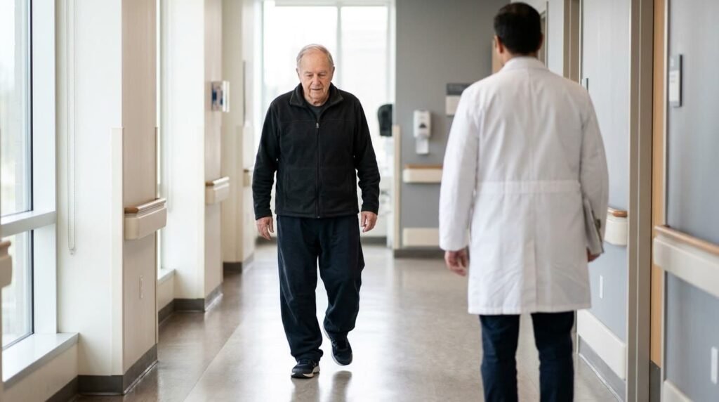
Observation starts with walking. An antalgic gait shortens the time spent on the painful leg. Standing inspection looks for bow legs or knock knees, which suggest more advanced wear. Both thighs get compared for muscle bulk. Wasting of the quadriceps suggests long-standing joint trouble.
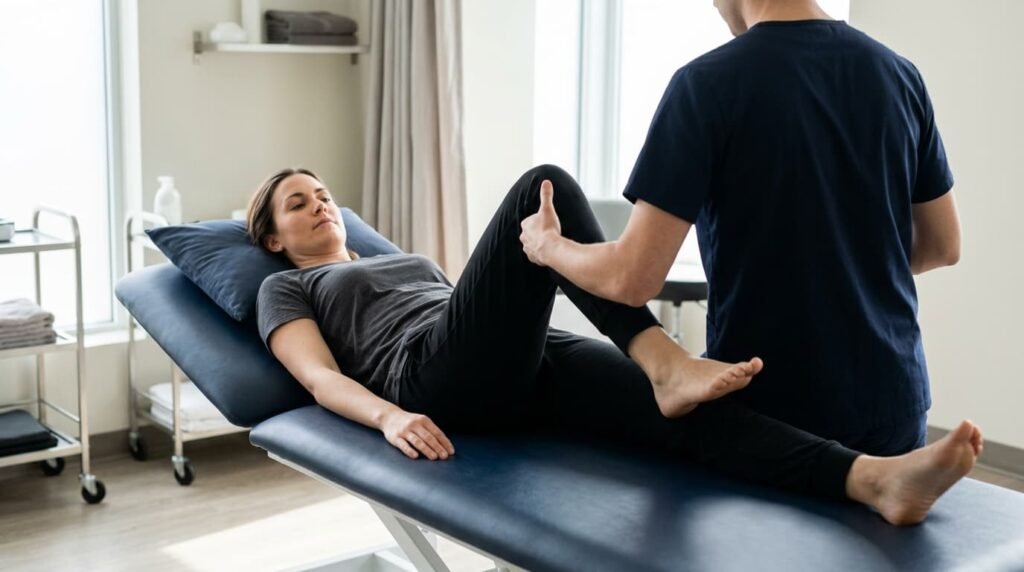
A structured physical examination of the knee then continues on the couch.
Palpation and Movement
With the patient lying down, the clinician presses along the joint lines. Tenderness suggests wear or meniscal irritation. Bone spurs in knee joints feel like hard bumps at the joint edge.
Range of movement comes next. Flexion beyond 130 degrees counts as normal for most people. Any flexion contracture is abnormal. The hip and lower back also need checking, because pain can refer to the knee from those areas.
Testing for Swelling and Fluid
The skin is checked for redness and warmth, and both knees are compared side by side. Water on the knee means excess fluid inside the joint. Three bedside tests detect it. The bulge sign suits small effusions. For moderate effusions, clinicians use the ballottement test method. The patellar tap test suits large effusions, where the kneecap taps the femur and bounces back.
A soft swelling behind the knee may be a Baker’s cyst. This cyst usually signals an underlying joint problem rather than causing pain itself.
What X-rays and MRI Scans Reveal
Imaging supports the diagnosis of knee osteoarthritis but rarely replaces the clinical picture. Radiography comes first. Further scans follow only when a specific question remains.
The Standing X-ray as the Gold Standard
A knee osteoarthritis x-ray works best with the patient standing, so the joint bears weight. Both knees are imaged for comparison. Standard views include front-to-back, side-on and a skyline view of the kneecaps.
Typical findings include joint space narrowing, bone spurs, cysts and misaligned joint surfaces. Subchondral sclerosis, a thickening of bone beneath the cartilage, often appears first.
A normal film does not rule out the condition. Early damage can hide on plain images, and pain does not always match the picture. NICE guidance allows a clinical osteoarthritis diagnosis without imaging in typical cases over 45. Typical means activity-related pain with morning stiffness under 30 minutes.
How Radiologists Grade the Film
Radiologists often use the Kellgren and Lawrence scale. It runs from grade 0, a healthy knee, to grade 4, severe disease. Grade 1 shows doubtful narrowing and possible small bone spurs. Grade 2 shows definite bone spurs and possible narrowing.
Some people with severe films cope well, and others with mild films struggle daily. Treatment therefore follows symptoms, not the picture alone.
When an MRI Adds Value
MRI for knee arthritis rarely helps in typical cases over 40. It shows soft tissue that plain films cannot, including cartilage, menisci and ligaments. A knee MRI scan becomes useful when the joint locks or gives out. It also helps when pain seems out of proportion to the X-ray.
MRI also detects bone marrow lesions, early cartilage loss and meniscal tears. Ultrasound shows fluid and soft tissue at the bedside. CT shows bone detail. These tools answer narrow questions rather than serve as routine tests.
Blood Tests, Joint Fluid and Red Flags
The diagnosis of knee osteoarthritis never relies on blood work. Osteoarthritis blood tests simply exclude rheumatoid arthritis, gout and other inflammatory causes. Joint fluid analysis checks for infection and crystals when the knee feels hot or swollen.

A hot, red, rapidly swelling knee needs urgent medical review. Gout in the knee and septic arthritis can both present this way. Fever, feeling unwell, or inability to bear weight raise the urgency further.
Several swollen joints, rash, weight loss or fatigue suggest an inflammatory condition. These signs justify specialist input.
Conditions That Mimic Osteoarthritis
Several problems copy the pain pattern. Patellofemoral pain syndrome causes front-of-knee pain on stairs, often in younger adults. Meniscal tears add catching and sharp pain with twisting. Hip disease can refer pain to the knee.
Rheumatoid arthritis usually affects several joints and brings prolonged morning stiffness. Careful examination separates these conditions in most cases. Imaging resolves the rest.
Diagnosis in Younger Adults and After Injury
Osteoarthritis is not limited to older age. Adults in their 30s and 40s can develop it, usually after an earlier injury. Common triggers include anterior cruciate ligament tears, meniscal surgery and fractures near the joint.
Why the Picture Looks Different
Younger knees often show pain with catching or swelling rather than steady aching. Standing films may look normal for years. Early cartilage damage can hide from plain images. MRI then plays a larger role, since it reveals cartilage and meniscal damage sooner.
What Clinicians Weigh
- Time since the original injury
- Whether the knee feels unstable
- Results of earlier scans or operations
- Work and sport demands
Early recognition matters most here, because decades of joint use lie ahead. Muscle strengthening and weight control protect the knee while longer-term plans take shape.
Who Treats Osteoarthritis and When to Refer
Care usually starts with a GP, who coordinates tests and early treatment. Physiotherapists guide exercise and joint protection. A rheumatologist reviews cases with inflammatory features. An orthopaedic surgeon assesses knees where pain control fails or movement drops sharply. Non-surgical knee specialists offer injection options between those stages.
When Referral Makes Sense
Referral to a rheumatologist suits cases with:
- Signs of inflammatory arthritis
- Swelling in several small or large joints
- Weight loss, fatigue or rash
Referral to an orthopaedic surgeon suits cases with:
- Pain that no longer responds to medication
- Mobility loss that disrupts daily tasks
- Failure of exercise and other conservative plans
What Follows a Confirmed Diagnosis
Treatment aims to ease pain, improve function and slow progression. Most plans combine several measures.

Exercise, Weight and Daily Habits
Exercise remains the first and most effective step. Cycling, swimming and walking strengthen the muscles that steady the joint. Think of the quadriceps as tent poles holding the structure upright.
Weight loss reduces load. Braces and shoe insoles shift pressure away from worn areas. Heat eases stiffness before activity. Cold calms swelling afterwards. Pacing helps too, since short walking bursts beat one long march. Small daily habits often outperform occasional heroic efforts.
Medicines and Injections
Paracetamol, anti-inflammatory tablets and topical creams manage flare-ups. Injections suit pain that persists despite those measures. Steroid injections give short-term relief. Hyaluronic acid injections add lubrication. Arthrosamid injection offers a longer-lasting hydrogel option for suitable patients, although it does not reverse joint damage.
Some people find acupuncture or massage helpful for short-term relief, although evidence remains mixed.
When Surgery Enters the Picture
Surgery remains for advanced cases. Options include osteotomy and partial or total knee replacement. Choice depends on age, pain intensity, disability and occupation.
Frequently Asked Questions
What does the diagnosis of knee osteoarthritis involve?
Clinicians start with a history and a hands-on examination. NICE guidance supports a clinical diagnosis without scans in many adults over 45. A standing X-ray follows when doubt remains. Further tests only exclude other conditions.
Can a knee osteoarthritis X-ray miss early disease?
Yes. Early cartilage changes often hide on plain films. Joint space narrowing appears later, after bone spurs in knee joints form. A normal film therefore does not exclude the condition.
When is MRI for knee arthritis worth having?
MRI helps when a knee locks, gives way or stays painful despite normal films. It shows cartilage, menisci and ligaments. Routine use in typical osteoarthritis adds little.
Does water on the knee always mean osteoarthritis?
No. Injury, infection, gout and inflammatory arthritis can all cause swelling. Bedside tests detect fluid, and aspiration identifies the cause when needed. A hot, red knee needs urgent review.
What do osteoarthritis blood tests show?
They do not confirm wear. Raised inflammatory markers or crystals point to another cause. Results guide referral to a rheumatologist when needed.
Who treats osteoarthritis if pain continues?
A GP usually leads early care and refers on when needed. Rheumatologists manage inflammatory features. Orthopaedic surgeons assess knees that fail simpler measures.
How long does a knee assessment take?
A first appointment usually lasts 10 to 20 minutes. Complex cases may need longer. Bringing earlier scans, letters and a medicine list speeds the process.
Do knock knees or bow legs raise the risk of osteoarthritis?
Yes. Misalignment shifts load to one side of the joint. Wear then develops faster on that side. Early assessment helps decide whether supports or other measures may slow progression.
Diagnosis of knee osteoarthritis combines careful questioning, hands-on testing and, where needed, a standing X-ray. Scans and blood tests mostly rule out other problems. Early recognition widens treatment choices and avoids unnecessary investigations.
Mr Syed Nadeem Abbas is a non-surgical knee and joint pain specialist who assesses suspected knee osteoarthritis.
Read more: Massage for knee pain eases stiffness and muscle tension when applied safely
Read more: Radiotherapy for Arthritis Calms Painful Joints Using Very Low Doses of Radiation


