Bone Marrow Lesion Knee Pain Points to a Deeper Joint Problem

✅ Medically reviewed | Updated August 2026
A bone marrow lesion knee finding often surprises patients expecting a straightforward diagnosis. An MRI scan can reveal a bright patch inside the subchondral bone. This happens long before pain fully explains itself. The finding is not a tumour, and it is not simple swelling. It is a zone of active bone change closely linked with knee osteoarthritis. Understanding what a bone marrow lesion means helps patients plan the right treatment path. That path might involve conservative care, targeted injections, or a more advanced procedure.
What Is a Bone Marrow Lesion in the Knee?
A bone marrow lesion (BML) sits within the subchondral bone. This is the dense layer directly beneath the articular cartilage. On fat-suppressed MRI sequences, such as STIR or fat-saturated T2, a BML shows up as a bright area. Earlier reports called this finding bone marrow oedema. That term rested on an assumption that fluid build-up explained the signal.
Later research shifted that view considerably. The bright signal reflects trabecular micro-damage, inflammatory activity, and abnormal bone remodelling. It does not reflect straightforward fluid accumulation. This distinction matters clinically: an active biological process behaves very differently from a passive pool of fluid.
Bone marrow lesion symptoms do not always match the size of a lesion on imaging. Some patients with large lesions report only mild discomfort. Others with small lesions experience considerable pain. Clinicians weigh MRI findings alongside physical examination before concluding severity.
Bone Marrow Lesion Causes
Several distinct pathways lead to a knee bone marrow lesion.
- Knee osteoarthritis – cartilage wear increases mechanical stress on the bone beneath it, triggering remodelling and lesion formation.
- Trauma – bone bruises, stress fractures, and ligament injuries such as ACL tears can produce acute subchondral oedema knee findings.
- Inflammatory arthritis – rheumatoid arthritis and related conditions drive immune-mediated bone changes.
- Metabolic factors – gout and other crystal-related conditions occasionally contribute to bone marrow abnormalities.
Clinical resources often grade these lesions by severity, from mild early changes through to advanced, extensive damage. Mild grades tend to follow minor trauma or early joint wear. Moderate grades often bring more noticeable pain and swelling. The most advanced grade usually accompanies significant osteoarthritis or major injury, and it typically needs a more intensive treatment plan. UK specialists lean on symptom correlation and MRI appearance together when deciding how urgently to act.
Subchondral Bone, Cartilage Loss, and Knee Osteoarthritis Treatment Decisions
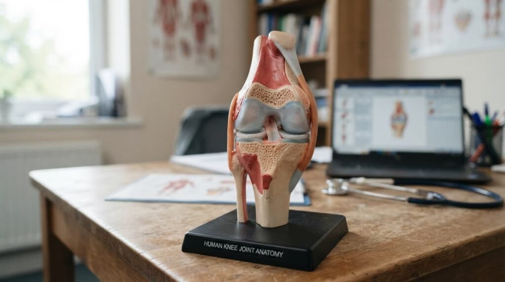
The subchondral bone and the cartilage above it behave as a single functional unit. Damage in one layer tends to influence the other over time. This bidirectional relationship reframes how a diagnosis should be read. A lesion is not an isolated bone finding. It forms part of a wider pattern affecting cartilage loss that knee patients often already notice through stiffness and swelling.
How Load Distribution Drives Cartilage Change
Established bone marrow lesions correlate with faster cartilage volume loss in longitudinal osteoarthritis studies. Abnormal load transmission across the joint appears central to this process. When forces concentrate unevenly, the bone beneath the cartilage absorbs that mechanical stress directly. Ongoing inflammation may then interfere with normal cartilage maintenance immediately above the lesion.
Why This Finding Deserves Clinical Attention
At a cellular level, mesenchymal stem cells within these lesions show measurable alterations in patients with osteoarthritis. Whether that change causes impaired cartilage repair, or simply reflects existing damage, remains debated among researchers. The practical takeaway is simple: this finding deserves proper clinical attention, not dismissal as an incidental MRI note.
Bone Marrow Lesion Knee Treatment Options
Treatment choice depends on lesion severity, symptom burden, and how far knee osteoarthritis has already progressed. Several distinct categories of bone marrow lesion treatment exist. Each works through a different mechanism.
Standard Intra-Articular Injection Approaches
Corticosteroid, hyaluronic acid, and PRP injection knee protocols remain widely used across UK knee clinics. Each carries a reasonable evidence base for short-to-medium-term pain relief and improved function. Their mechanism operates within the joint cavity and synovium. It does not reach inside the bone itself.
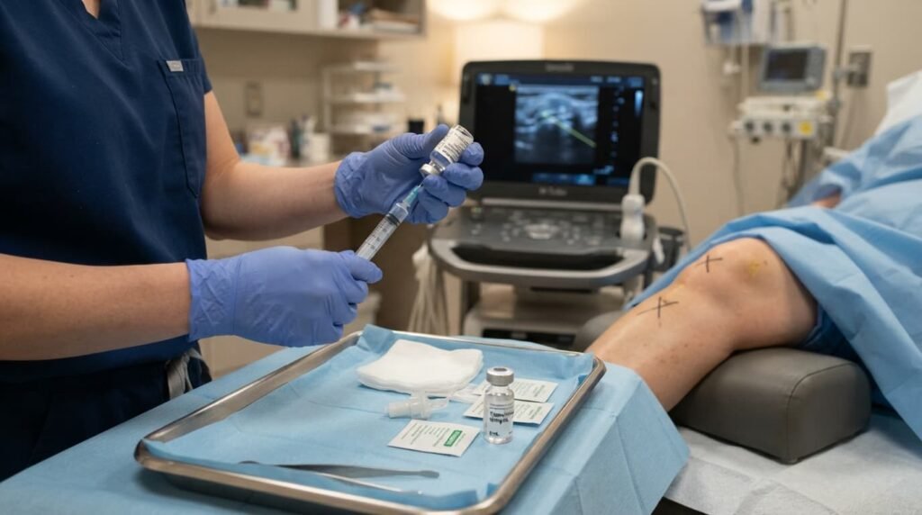
- Corticosteroids reduce intra-articular inflammation.
- Hyaluronic acid restores some of the viscous quality synovial fluid loses in osteoarthritis.
- PRP delivers concentrated growth factors that modulate the local inflammatory environment.
None of these three agents was designed with lesion volume reduction as a primary goal. Current evidence does not document a direct structural effect on subchondral pathology from any of them. That does not diminish their value for pain control; it simply narrows their therapeutic scope. PRP Arthrosamid outcomes diverge mainly because one relies on biology, the other on physical integration.
Polyacrylamide Hydrogel, Arthrosamid, and iPAAG
Polyacrylamide hydrogel knee treatment, marketed in the UK as Arthrosamid, behaves differently from the agents above. Rather than dispersing through joint fluid, it integrates permanently into the synovial membrane as a non-degradable scaffold. It is composed of roughly 2.5% cross-linked polyacrylamide and 97.5% water. It functions as a mechanical buffer that may redistribute load across the bone beneath the cartilage.
That structural integration makes this treatment mechanistically interesting for subchondral pathology, beyond simple symptom control. A 2022 study reported measurable reductions in patellofemoral bone marrow lesions after a single iPAAG injection. Patients in that study had advanced knee osteoarthritis. The finding is preliminary, drawn from a single-centre, non-randomised design. It remains, even so, the clearest available citation linking an injection directly to reduced lesion burden.
Separate research involving 269 patients and 314 knees confirmed sustained improvements in pain and function over two years. Systematic lesion measurement was not a primary endpoint in that larger cohort, though the functional results were consistent. This Arthrosamid treatment follows a consultation, imaging review, and a single outpatient procedure in most cases.
Sub-Chondroplasty and Bone Marrow Concentrate Injection
Sub-chondroplasty and bone marrow concentrate injection occupy a distinct category from the approaches above. These are direct, bone-targeting procedures rather than joint-cavity injections.
- Sub-chondroplasty – the needle enters bone directly, not the joint cavity. Performed under X-ray guidance, a flowable calcium phosphate substitute fills the lesion itself, providing scaffolding for new bone growth. It is typically combined with arthroscopy, so cartilage defects can be addressed in the same session.
- Bone marrow concentrate injection – delivers a patient’s own concentrated cells into the lesion under image guidance.
- Core decompression surgery – an older surgical technique that relieves pressure on the affected bone and promotes blood flow. It now sits behind less invasive options for many patients.
These direct approaches generally suit patients whose symptoms have not responded to conservative management. They are rarely offered as a first-line choice, given the surgical access and recovery time involved.
Diagnosing a Knee Bone Marrow Lesion
MRI and Imaging Findings
MRI remains the gold-standard tool for detecting a bone marrow lesion in the knee. It offers detailed images of trabecular changes long before X-rays would show anything abnormal. Fat-suppressed sequences highlight the bright signal characteristic of active lesions. Adjacent structures, such as cartilage and ligaments, can be assessed within the same scan.

Related findings sometimes appear alongside a knee MRI bone marrow lesion result. These include osteochondritis dissecans, meniscal changes, and early cartilage thinning. X-rays remain useful for assessing joint space narrowing and bone spurs. They lack, however, the sensitivity to detect subchondral oedema knee changes directly.
Physical Examination and Symptom History
Physical examination adds another layer of information. A clinician assesses range of motion, swelling, tenderness, and joint stability by hand. Symptom history matters just as much as imaging. A patient’s description of when pain worsens and what eases it guides how the MRI findings should be interpreted. Physical examination, symptom history, and imaging together build a complete clinical picture. No single test tells the full story on its own.
Living With Knee Joint Inflammation and Planning Next Steps
Recognising Early Signs
Persistent knee joint inflammation linked to a bone marrow lesion can restrict daily activity well before structural damage becomes severe. Stiffness after rest, swelling around the joint line, and reduced range of motion are common early signs. These signs deserve prompt reporting rather than silent management, since earlier assessment tends to preserve more treatment options.
Interventional orthobiologics and stem cell therapy knee research continue to expand the non-surgical toolkit available to patients. Video explainers on non-surgical joint pain and arthritis treatment sit on the Dr SNA Clinic YouTube channel. They offer a useful visual companion to written information for anyone weighing up these options. This Arthrosamid injection in London typically starts with an MRI-based assessment to confirm suitability before any procedure date is set.
When to Watch and When to Act
Not every finding requires immediate intervention. Size, location, symptom severity, and response to conservative measures all influence whether watchful waiting or active treatment makes sense. Physiotherapy and load management often form the first practical step for milder cases. That judgement cannot be made from an MRI report alone; it needs a proper clinical conversation.
Daily Habits That Support Joint Health
Simple daily habits can also support joint health alongside formal treatment. Maintaining a healthy weight reduces mechanical load passing through the knee with every step. Low-impact activity, such as swimming or cycling, keeps surrounding muscles strong without adding excessive joint stress. Patients recovering from a knee procedure often benefit from broader joint-care guidance alongside lesion-specific advice. The two conditions frequently overlap in practice.

How Related Terms Connect to a Bone Marrow Lesion Knee Diagnosis
Several terms discussed above describe different pieces of the same clinical picture. Bone marrow oedema and bone marrow lesion describe the same underlying MRI finding. The newer term reflects updated understanding of the inflammatory process involved. Subchondral bone marrow lesion is simply a more formal version of the same term. Both point to the dense bone layer beneath the cartilage, the specific site where lesions form.
Cartilage degeneration knee changes and cartilage loss knee symptoms represent the downstream consequence of an unmanaged lesion. This reflects the bidirectional relationship between these two tissue layers. Corticosteroid injection knee therapy, hyaluronic acid injection knee treatment, and PRP injection knee protocols address joint-level symptoms. None were designed to shrink subchondral lesions directly.
Polyacrylamide hydrogel knee therapy, Arthrosamid, and iPAAG injection describe the same treatment viewed from three angles: material composition, UK brand name, and clinical shorthand. Both direct-delivery bone procedures target the lesion itself rather than the joint space, unlike standard injections. A knee bone cyst can develop when a lesion progresses untreated. Earlier assessment helps avoid that long-term consequence.
Frequently Asked Questions
What is a bone marrow lesion in the knee?
A bone marrow lesion is an area of trabecular micro-damage and inflammatory change within the subchondral bone. It sits just beneath the cartilage. It typically appears as a bright patch on fat-suppressed MRI sequences. The term replaced bone marrow oedema once research clarified that inflammation, rather than simple fluid, drives the signal.
What are the main bone marrow lesion symptoms to watch for?
Bone marrow lesion symptoms include localised knee pain, stiffness after rest, and swelling around the joint line. Symptom severity does not always match lesion size on MRI. A specialist assessment helps determine whether findings warrant active treatment or ongoing monitoring.
Can standard injections treat a bone marrow lesion?
Standard corticosteroid, hyaluronic acid, and PRP injection knee protocols act mainly within the joint cavity rather than the bone. Current evidence does not confirm they reduce lesion volume directly. They can, however, still relieve associated joint pain effectively.
How does polyacrylamide hydrogel differ from other knee injections?
Polyacrylamide hydrogel, known in the UK as Arthrosamid, integrates permanently into the synovial membrane rather than dispersing through joint fluid. This iPAAG injection may redistribute mechanical load across the bone beneath the cartilage. That offers a plausible pathway toward reduced lesion burden over time.
When is subchondroplasty considered for a knee bone marrow lesion?
Subchondroplasty is generally reserved for symptomatic, imaging-confirmed lesions that have not responded to conservative management or standard injection. It involves delivering a bone substitute directly into the lesion under X-ray guidance, often alongside arthroscopy.
Do bone marrow lesions always lead to knee osteoarthritis?
Not every lesion progresses to advanced osteoarthritis, though the two conditions frequently occur together. Early assessment and appropriate treatment can reduce the likelihood of rapid progression. Regular monitoring remains sensible for anyone with a confirmed finding on MRI.
Mr Syed Nadeem Abbas advises patients with persistent knee symptoms to seek thorough specialist assessment, not imaging alone.
Read more: How Vitamins and Joint Pain Are Connected: A Complete Guide
Read more: Knee Arthritis Treatment Options: A Complete Guide to Managing Knee Osteoarthritis