Synovial Fluid in Knee Explained With Its Causes and Treatment

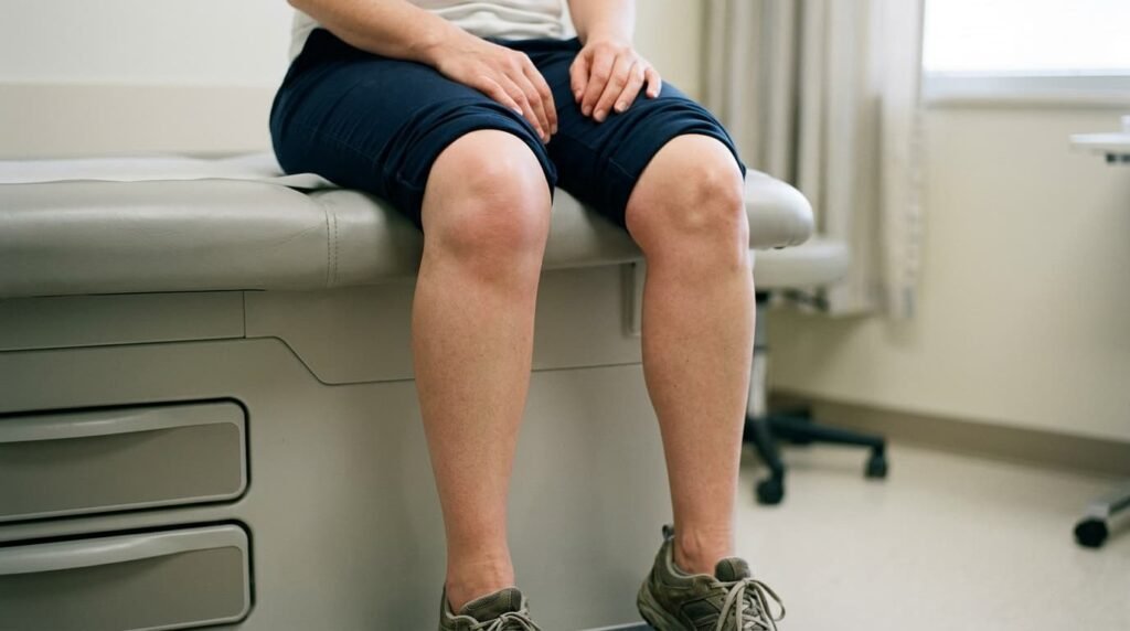
Synovial fluid in knee joints does more than most people think. It cushions the joint, feeds the cartilage, and keeps movement smooth every time the leg bends, walks, or climbs stairs. A small amount sits inside a healthy knee at all times, renewed constantly by cells lining the joint capsule. Problems start when too much builds up, or when its colour, thickness, or cell content changes in response to injury, wear, or disease. These shifts often show up as visible swelling, stiffness, or pain that does not settle with rest. Doctors rely on a close look at this fluid, through aspiration and lab analysis, to work out exactly what is going wrong inside the joint.
What Is Synovial Fluid and Why the Knee Needs It
Every joint relies on a thin layer of fluid for smooth movement. The knee is no different.
Function of Synovial Fluid in Joint Movement
The main synovial fluid function is to cut friction between the bones during walking or bending. This thick, slightly sticky fluid also feeds the cartilage, which has no blood supply of its own. Without enough fluid, joint surfaces would grind against each other. That grinding speeds up wear and tear over time. Special cells lining the joint capsule make the fluid. They keep renewing it in small amounts. The fluid also carries away waste from cartilage cells. In a healthy knee, only a small amount is present. It sits just thick enough to coat the joint surfaces.
Synovial Fluid Colour Normal Range and Appearance
A synovial fluid colour normal reading is pale yellow and clear. The fluid often stretches slightly between two fingers, a simple test doctors sometimes use. Cloudy, thick, or watery fluid can be an early clue that something has changed. A shift to red or brown often points to bleeding inside the joint. A more opaque, pus-like look can suggest infection. These visual checks usually come first, before any lab testing begins.
What Causes Fluid on the Knee
Several conditions can trigger a build-up of fluid. The cause often shapes the treatment plan that follows.
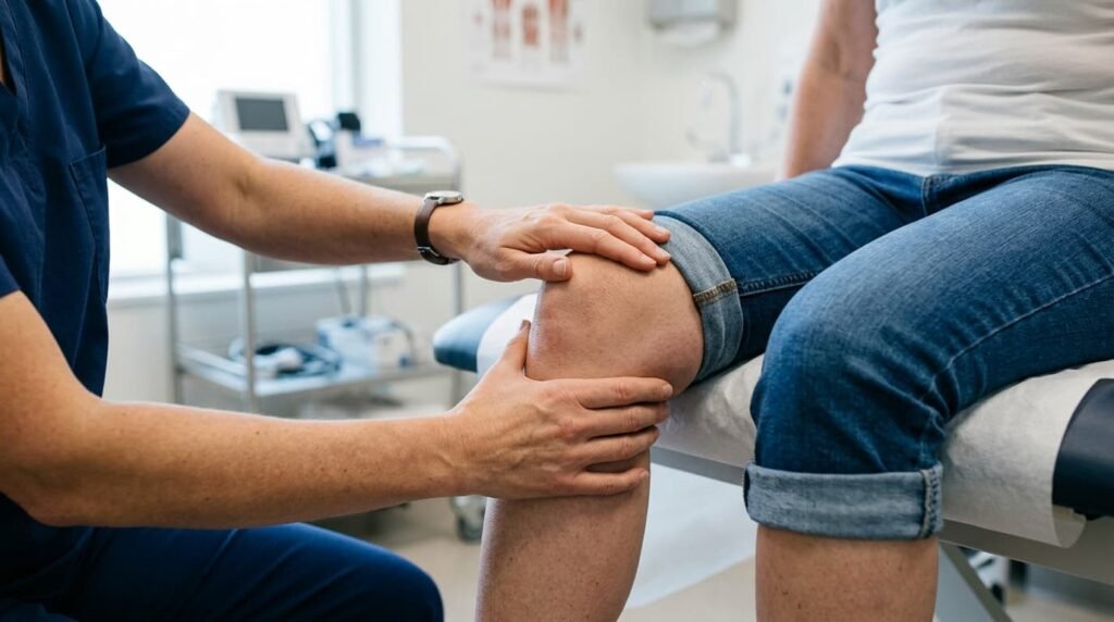
Common Causes of Swollen Knee
A swollen knee causes a clear change in shape. It can appear suddenly after an injury, or build up slowly with long-term conditions. A fall or sports injury often leads to fast swelling within hours. Repeated strain from overuse can also cause a slow build-up over weeks. Infection is less common, but it brings swelling along with warmth and fever. Noting how fast the swelling appeared helps narrow down the likely cause.
Osteoarthritis and Joint Fluid Changes
Osteoarthritis joint fluid often increases as cartilage wears down over time. The joint reacts to this wear by producing more fluid than usual. This is the most common reason older adults notice fluid on the knee causes linked to mechanical stress rather than sudden injury. The fluid itself may turn slightly thinner than normal. It loses some of its natural cushioning quality. Pain tends to get worse with activity and ease with rest. This pattern sets it apart from inflammatory causes.
Rheumatoid Arthritis Joint Fluid Test Findings
A rheumatoid arthritis joint fluid test usually shows a much higher white cell count than osteoarthritis. This points to active inflammation rather than simple wear. The joint lining becomes thickened and overactive. It then produces extra fluid as part of the immune response. Both knees are often affected at the same time. This differs from the one-sided pattern seen with mechanical causes. Morning stiffness lasting more than thirty minutes often comes with this condition.
Joint Effusion Symptoms to Watch For
Catching the right symptoms early can stop a minor issue from turning into something more serious.
Signs That Need Medical Attention
Joint effusion symptoms usually include visible swelling, tightness, and less range of motion than the other knee. Warmth over the joint, combined with redness, raises concern about infection. This combination needs urgent checking. A fever alongside knee swelling should never be ignored. Septic arthritis can damage cartilage fast if left untreated. Other warning signs include sudden trouble bearing weight, a locking joint, or fluid that keeps coming back after drainage. Any of these call for a professional opinion without delay.
Synovial Fluid Analysis Explained
Once fluid is collected, lab analysis gives the clearest picture of what is happening inside the joint.
What Is Synovial Fluid Analysis Used For
Synovial fluid analysis helps tell apart mechanical wear, inflammatory disease, infection, and bleeding disorders. Each one needs a different treatment path. The sample gets checked for appearance, thickness, and clarity first. It then moves on to more detailed chemical and microscopic tests. Glucose and protein levels in the fluid get compared with blood values. A large gap between the two can signal infection. This mix of checks makes the test one of the more reliable tools for joint problems.
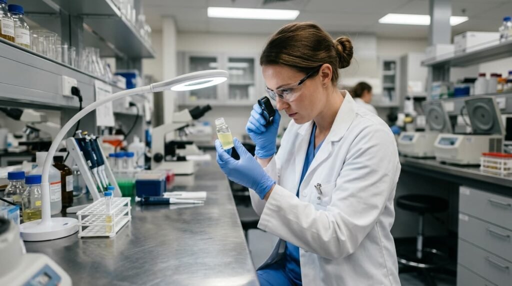
Synovial Fluid Analysis Results and What They Mean
Synovial fluid analysis results usually fall into groups based on white blood cell counts. These range from non-inflammatory to clearly infectious. A low count under roughly 200 cells points to a mechanical issue rather than active inflammation. Counts climbing into the thousands suggest an inflammatory process, such as gout or rheumatoid disease. A count far higher than that, often paired with visible pus, raises strong concern for septic arthritis. That finding needs treatment right away.
Inflammatory vs Non-Inflammatory Synovial Fluid
The inflammatory vs non-inflammatory synovial fluid split guides much of the early decision-making. This happens before more specific test results come back. Non-inflammatory fluid tends to stay clear and pale. It also shows a lower cell count and little change in thickness. Inflammatory fluid is often cloudier and thinner. It carries a higher share of certain white cells. This one classification step can rule out several causes early. It narrows down what further testing is actually needed.
Gout, Pseudogout, and Crystal Analysis
Crystal-related joint disease leaves a clear fingerprint that lab analysis can usually spot with confidence.
Gout vs Pseudogout Symptoms
Gout vs pseudogout symptoms can look much the same at first. Both cause sudden, sharp joint pain and swelling. Gout classically affects the big toe, but it can also strike the knee. Diet and dehydration often trigger a flare. Pseudogout tends to favour larger joints, including the knee. It is more common in older adults. Both conditions can flare without warning and settle within days. They often return later if left untreated.
Synovial Fluid Gout Findings
Synovial fluid gout cases show needle-shaped crystals under polarised light. This finding confirms the diagnosis almost right away. Pseudogout instead produces crystals shaped like rhombi, with a different optical pattern. This lets the two conditions get told apart reliably. Crystal identification is one of the most useful parts of the whole testing process. Treatment differs between the two conditions, even though the symptoms look similar.
The Knee Aspiration Procedure
Removing fluid from the knee is a fairly quick procedure. It can serve both diagnosis and treatment at the same time.
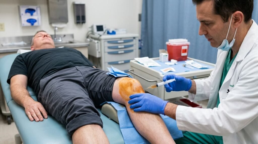
What Happens During Arthrocentesis Knee Procedure
An arthrocentesis knee procedure starts with cleaning the skin well to cut infection risk. A local numbing agent is used next. Most people feel pressure rather than sharp pain during the process. The clinician then guides a needle into the joint space. They draw out the fluid using a syringe. Ultrasound guidance is sometimes used to improve accuracy. This is especially true when the knee is less swollen than usual.
Joint Fluid Aspiration Step by Step
Joint fluid aspiration usually takes only a few minutes from start to finish. This makes it a simple outpatient procedure. Once enough fluid comes out, it gets moved into sterile containers for lab testing right away. A small dressing covers the entry point. Most people can walk out shortly after. This simple process is part of why aspiration stays a first step for unexplained knee swelling.
How Is Joint Fluid Removed and Pain Relief
How is joint fluid removed comes down to a needle and syringe guided into the swollen joint space. Knee joint fluid removal often brings relief almost right away, as pressure inside the joint drops. Many people say they can bend the knee more freely within hours of the procedure. This benefit can be short-lived if the root cause of the build-up is not treated separately. For that reason, aspiration is often paired with further checks rather than used alone.
Knee Joint Aspiration Recovery and Drainage Risks
Recovery is usually simple. A few precautions help avoid problems in the days that follow.
Knee Fluid Drainage Risks
Knee fluid drainage risks are generally low. Mild soreness and bruising near the injection site are common for a day or two. Fluid building back up is the most common issue reported. This is more likely if the original cause stays active. Rare but more serious risks include infection from the procedure or, very rarely, damage to nearby cartilage. Picking an experienced clinician under clean conditions keeps these risks low.
Aftercare Advice
Knee joint aspiration recovery usually means resting the leg, using ice, and skipping hard activity for a short time. A compression bandage can help stop fluid from building back up too fast. Any growing redness, warmth, or fever in the days after aspiration should get reported right away. These can be signs of infection. Most people get back to normal activity within a few days. This depends on what caused the swelling in the first place.
Knee Effusion Treatment Options
Once the cause is clear, treatment can be matched to whether the problem is mechanical, inflammatory, or infectious.
Water on the Knee NHS Pathway
The water on the knee NHS pathway usually starts with a GP check. A referral to a specialist follows if the cause stays unclear. Early NHS care often focuses on rest, anti-inflammatory medicine, and watching the joint before trying anything more invasive. Wait times for specialist scans or rheumatology review can vary a lot by area. Knee effusion treatment under this route tends to follow a step-by-step approach. It only moves forward when simpler steps fail.
When Private Specialist Treatment May Help
For people facing long waits, or wanting a faster and more focused check, private clinics offer quicker access to scans, aspiration, and targeted injections. This route can help a great deal when swelling keeps returning and disrupts daily life, especially when no clear answer has come from standard channels yet. Treatment plans in this setting often get built around the specific findings from synovial fluid analysis rather than a one-size-fits-all protocol.
Frequently Asked Questions
What does synovial fluid analysis check for?
Synovial fluid analysis checks the colour, thickness, and cell content of joint fluid. This helps spot infection, inflammation, gout, or mechanical wear in the knee. The fluid sample also goes through chemical testing, comparing glucose and protein levels against blood values to flag possible infection. Together, these checks let doctors choose the right treatment path much faster than relying on symptoms alone.
What is synovial fluid and why does the knee produce it?
What is synovial fluid comes down to a natural lubricant made inside the joint capsule. It cuts friction and feeds cartilage during movement. The fluid also carries away waste produced by cartilage cells, since cartilage has no blood supply of its own to manage this directly. Only a small amount sits in a healthy knee, just enough to keep the joint surfaces gliding smoothly.
What is the main fluid on the knee?
Fluid on the knee causes range from direct injury and overuse to osteoarthritis, rheumatoid arthritis, gout, and sometimes infection. Sudden swelling after a fall or sports injury usually points to trauma, while a slower build-up over weeks often signals a long-term condition like osteoarthritis. Noting how quickly the swelling appeared, and whether it comes with warmth or fever, helps narrow down the likely cause before any testing begins.
How is the knee aspiration procedure performed?
The knee aspiration procedure involves cleaning the skin, numbing the area, and using a needle to draw out extra fluid for relief and testing. The whole process usually takes only a few minutes, and most people feel pressure rather than sharp pain throughout. A small dressing is placed over the entry point afterwards, and most people are able to walk out shortly after the procedure finishes.
Is knee joint aspiration recovery painful?
Knee joint aspiration recovery is usually mild. It brings slight soreness for a day or two, and most people return to normal activity quickly. Resting the leg, using ice, and wearing a compression bandage for a short time can help stop fluid from building back up too fast. Any growing redness, warmth, or fever in the days that follow should be reported promptly, since these can signal infection.
What is the difference between gout and pseudogout symptoms?
Gout vs pseudogout symptoms both bring sudden joint pain and swelling. They come from different crystal types, confirmed through fluid analysis. Gout classically affects the big toe but can also strike the knee, often triggered by diet or dehydration, while pseudogout tends to favour larger joints and is more common in older adults. Lab testing under polarised light is the most reliable way to tell the two apart, since the symptoms alone can look almost identical.
What treatment is available through the water on the knee NHS route?
The water on the knee NHS pathway usually starts with a GP check. It may move on to specialist referral, aspiration, or scans depending on how severe things are. Early NHS care tends to focus on rest, anti-inflammatory medicine, and monitoring before more invasive options come into play. Wait times for specialist review can vary by area, which is why some people look at private clinics for faster access to testing and treatment.
Synovial fluid in knee joints does far more than lubricate movement. Changes in its amount or makeup often point to a problem worth checking. From osteoarthritis to gout and infection, the causes vary widely. Accurate testing through aspiration and lab analysis remains the most reliable way to reach a clear diagnosis and a fitting treatment plan.
Read more: Diabetic Knee Pain and the Treatments That Actually Help
Read more: How Ultrasound-Guided Knee Injections Improve Pain Relief